43 spinal cord model with labels
Spinal Cord Stimulation and Training - ClinicalTrials.gov This study will help the investigators better understand the changes in short-term excitability and long-term plasticity of corticospinal, reticulospinal and spinal neural circuits and how the changes impact the improvements of spinal cord stimulation (SCS) mediated motor function. Detailed Description: What are the 12 cranial nerves? Functions and diagram - Medical News Today Scientists use Roman numerals from I to XII to label the cranial nerves in the brain. The 12 cranial nerves include the: olfactory nerve optic nerve oculomotor nerve trochlear nerve trigeminal...
faseb.onlinelibrary.wiley.com › journal › 15306860The FASEB Journal - Wiley Online Library The FASEB Journal publishes high quality and impactful multidisciplinary research covering biology and biomedical sciences at every level of organization: atomic, molecular, cell, tissue, organ, organismic, and population.

Spinal cord model with labels
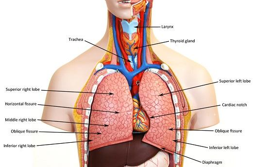
Human torso model labeled organs | labquiz Labeled human torso models feature clear views of the vertebrae spinal cord spinal nerves vertebral arteries lungs stomach liver intestinal tract kidneys heart and more. In Stock Ships Out Within 24 Hours This Anatomy Toy is a visual representation of the human torso and its internal organs. The abdomen and thorax are two regions of the torso. Ubap1 knock-in mice reproduced the phenotype of SPG80 A Confocal images of spinal cord sections from 7-month-old mice stained with ubiquitin (red), LC3 (green), and DAPI (blue). Yellow in the merged images indicates colocalization of ubiquitin and LC3. › articles › s41597/021/00941-8Open-access quantitative MRI data of the spinal cord and ... Aug 16, 2021 · In a companion paper by Cohen-Adad et al. we introduce the spine generic quantitative MRI protocol that provides valuable metrics for assessing spinal cord macrostructural and microstructural ...
Spinal cord model with labels. › pmc › articlesNovel stochastic framework for automatic segmentation of ... Animal studies showed that spinal cord transection reduced muscle mass of hind-limb extensors between 20% and 40% in one month [2–4]. Individuals with chronic SCI also showed cross-sectional area of the whole thigh, knee extensors and plantar flexors that were about 30% smaller compared to non-disabled individuals [ 5 , 6 ]. Mapping of neuroinflammation-induced hypoxia in the spinal cord using ... In our present study, we investigated the changes in oxygenation and analyzed the vascular perfusion of the spinal cord in a rodent model of MS. We performed multispectral optoacoustic tomography of the lumbar spinal cord before and after an oxygen enhancement challenge in mice with experimental autoimmune encephalomyelitis (EAE), a model for MS. Anatomy, Back, Spinal Meninges - StatPearls - NCBI Bookshelf The spinal cord and brain are encased within three layers of tissue called the meninges. The spinal meninges specifically enclose the spinal cord and stretch from the brainstem down to the filum terminale. The layers of the meninges are, from deep to superficial, the pia mater, the arachnoid mater, and the dura mater. The names of these layers give information regarding their qualities. Pia ... Learn the spinal cord with diagrams and quizzes | Kenhub Here you'll simply fill in the blanks with the name of the structure corresponding to the diagram label. You can also download the spinal cord diagram labeled if you'd like to scribble and make notes before attempting the labeling activity. DOWNLOAD PDF WORSHEET (BLANK) DOWNLOAD PDF WORKSHEET (LABELED) Accelerate your learning with quizzes
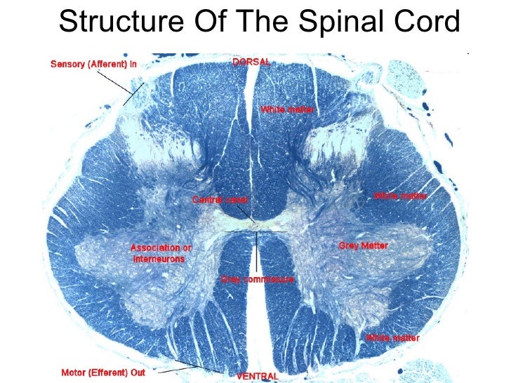
Spinal Cord Stimulation in Spinal Muscular Atrophy (SCSinSMA) Intervention Model Description: Single-arm, open-label study performed to quantify variables that are predictive of the efficacy of spinal cord stimulation to improve motor control. Masking: ... Spinal cord stimulation produces tingling sensations and other type of sensory phenomena. It is important to document that stimulation intensities ... Spinal cord: Anatomy, structure, tracts and function | Kenhub The spinal cord is made of gray and white matter just like other parts of the CNS. It shows four surfaces: anterior, posterior, and two lateral. They feature fissures (anterior) and sulci (anterolateral, posterolateral, and posterior). The gray matter is the butterfly-shaped central part of the spinal cord and is comprised of neuronal cell bodies. Spinal Cord Stimulation Therapy for Hereditary Spastic Paraplegias ... It's a single-center, prospective, open label clinical study with a 12 months follow-up period, to investigate the therapeutic effect and safety of spinal cord stimulation (SCS) on motor function and gait in patients with pure Hereditary Spastic Paraplegias. Detailed Description: Procedure Bstructure Of The Spinal Cord - GUWS Medical 1. Review a textbook section on the spinal cord. 2. As a review activity, label figures 27.1, 27.2, and 27.3. 3. Complete Parts B and C of the laboratory report. 4. Obtain a prepared microscope slide of a spinal cord cross section. Use the low power of the microscope to locate the following features:
› types-of-spinal-cord-injuriesSpinal Cord Injury | Types of Spinal Cord Injuries ... 88 percent of spinal cord injury survivors who were single at the time of the accident are single five years later, compared to 65 percent in the general population. Two-thirds of sports-related spinal cord injuries are from diving, making it the most dangerous sport for the brain and spinal cord. Blank ear diagrams and quizzes: The fastest way to learn | Kenhub It helps you to memorize the names and their locations, which in turn will aid you to remember their functions. Below, you can download both the blank ear diagram to make some notes, and then try labeling the ear using the unlabeled ear diagram. Good luck! DOWNLOAD PDF WORKSHEET (BLANK) DOWNLOAD PDF WORKSHEET (LABELED) byjus.com › biology › spinal-cord-diagramSpinal Cord Diagram with Detailed Illustrations and Clear Labels The spinal cord is one of the most important structures in the human body. In fact, it is the most important structure for any vertebrates. Anatomically, the spinal cord is made up is made up of nervous tissue and is integrated into the spinal column of the backbone. Main Article: Spinal Cord – Anatomy, Structure, Function, and Spinal Cord Nerves Ventral Cord Syndrome - StatPearls - NCBI Bookshelf Ventral cord syndrome (VCS), also referred to as anterior cord syndrome or anterior spinal artery syndrome, is caused by any condition that leads to infarction of the ventral two-thirds of the spinal cord. Estimates for the incidence and prevalence of ventral cord syndrome vary, yet it is the most common type of spinal cord infarction. Patients with ventral cord syndrome present with ...
Print Activity 5: Examining the Human Torso Model flashcards | Easy Notecards
link.springer.com › article › 10Spatiotemporal Dynamics of the Molecular Expression Pattern ... Jul 05, 2022 · Nerve regeneration in adult mammalian spinal cord is poor because of the lack of intrinsic regeneration of neurons and extrinsic factors – the glial scar is triggered by injury and inhibits or promotes regeneration. Recent technological advances in spatial transcriptomics (ST) provide a unique opportunity to decipher most genes systematically throughout scar formation, which remains poorly ...

Pin on Histology - Spinal Cord and Ganglion
Spine anatomy diagrams and interactive vertebrae quizzes | Kenhub The spine diagram below highlights all of the vertebrae labeled. You can see the cervical vertebrae labeled at the top, the thoracic vertebrae labeled in the middle and the lumbar vertebrae labeled towards the bottom. Labeled overview of the vertebral column.

Spinal cord Jeopardy Review Game Answer Key
Spinal Cord Injury Neuroprotection With Glyburide (SCING) This neurological exam assesses sensory and motor impairments using touch and pin prick evaluations in each dermatome and muscle strength. Right and left body sides are evaluated independently. Lower the scores indicate greater degree of impairment. Touch and pin prick perception is scored using a 0-2 and NT grade.

Spinal cord - Neuroanatomy An Illustrated Colour Text, 4 ed.
Schwann Cell Anatomy - Human Anatomy - GUWS Medical Figure 25.1 Label this diagram of a motor neuron. Figure 25.2 Label the features of the myelinated nerve fiber. Figure 25.3 Micrograph of a multipolar neuron and neuroglia from a spinal cord smear (100x micrograph enlarged to 600x). -Nerve fiber (axon) general name for processes (either dendrites or axon) of the neuron.

Kidney Model – Human Body Help
FREE Human Body Systems Labeling with Answer Sheets - Homeschool Giveaways The free skeletal system labeling sheet includes a fill-in-the-blanks labeling of the main bones on the body. The free respiratory system labeling sheet includes a blank diagram to fill in the trachea, bronchi, lungs, and larynx. The free nervous system labeling sheet includes blanks to label parts of the brain, spinal cord, ganglion, and nerves.

💠Vertebral column. Spinal Anatomy. 2019-02-13
› articles › 326536How to tighten loose skin: 6 tips - Medical News Today Oct 03, 2019 · The effect of oral collagen peptide supplementation on skin moisture and the dermal collagen network: Evidence from an ex vivo model and randomized, placebo‐controlled clinical trials. https ...
Anatomy of spinal cord
These Are the 12 Cranial Nerves and Their Functions - Healthline Your cranial nerves are pairs of nerves that connect your brain to different parts of your head, neck, and trunk. There are 12 of them, each named for its function or structure. Their functions are...
Post a Comment for "43 spinal cord model with labels"