39 simple microscope diagram with labels
Label the microscope — Science Learning Hub Use this with the Microscope parts activity to help students identify and label the main parts of a microscope and then describe their functions. Drag and drop the text labels onto the microscope diagram. If you want to redo an answer, click on the box and the answer will go back to the top so you can move it to another box. Parts of a Microscope Labeling Activity - Storyboard That Create a poster that labels the parts of a microscope and includes descriptions of what each part does. Click "Start Assignment". Use a landscape poster layout (large or small). Search for a diagram of a microscope. Using arrows and textables label each part of the microscope and describe its function. Copy This Storyboard* More options
Simple Squamous Epithelium under a Microscope with a Labeled Diagram ... Simple columnar epithelium labeled. This is a labeled diagram of a simple columnar epithelium under a light microscope. I tried to show you both ciliated and nonciliated simple columnar epithelium. These diagrams show the cilia on the cell surface, rectangular cell, and elongated nucleus.

Simple microscope diagram with labels
Label the Microscope Diagram | Download Scientific Diagram - ResearchGate Download scientific diagram | Label the Microscope Diagram from publication: Laboratory Exercises in Microbiology: Discovering the Unseen World through Hands-on Investigation | Microbiology ... Simple Microscope - Parts, Functions, Diagram and Labelling Simple Microscope - Parts, Functions, Diagram and Labelling By Editorial Team March 7, 2022 A microscope is one of the commonly used equipment in a laboratory setting. A microscope is an optical instrument used to magnify an image of a tiny object; objects that are not visible to the human eyes. Table of Contents Simple Microscope: Definition, Principle, Parts, And Uses A simple microscope is a rudimentary magnification device that is capable of visibly enlarging small objects, so they can be viewed and studied in better detail. It was invented in the late 16th century, and is still being widely used today. Simple microscopes have a wide range of applications in various fields.
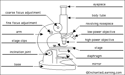
Simple microscope diagram with labels. Fluorescence Resonance Energy Transfer (FRET) Microscopy Presented in Figure 3 is a Jablonski diagram illustrating the coupled transitions involved between the donor emission and acceptor absorbance in fluorescence resonance energy transfer. Absorption and emission transitions are represented by straight vertical arrows (green and red, respectively), while vibrational relaxation is indicated by wavy yellow arrows. Microscope With Labels clip art | Microscope parts, Scientific method ... Microscope With Labels clip art | Microscope parts, Scientific method, Science diagrams From clker.com vector clip art online, royalty free & public domain Download Clker's Microscope With Labels clip art and related images now. Multiple sizes and related images are all free on Clker.com. D Dixie Tsutsaeva 2k followers More information Compound Microscope Parts, Functions, and Labeled Diagram The total magnification of a compound microscope is calculated by multiplying the objective lens magnification by the eyepiece magnification level. So, a compound microscope with a 10x eyepiece magnification looking through the 40x objective lens has a total magnification of 400x (10 x 40). Fluorescence - Wikipedia Fluorescence is the emission of light by a substance that has absorbed light or other electromagnetic radiation.It is a form of luminescence.In most cases, the emitted light has a longer wavelength, and therefore a lower photon energy, than the absorbed radiation.A perceptible example of fluorescence occurs when the absorbed radiation is in the ultraviolet region of the …
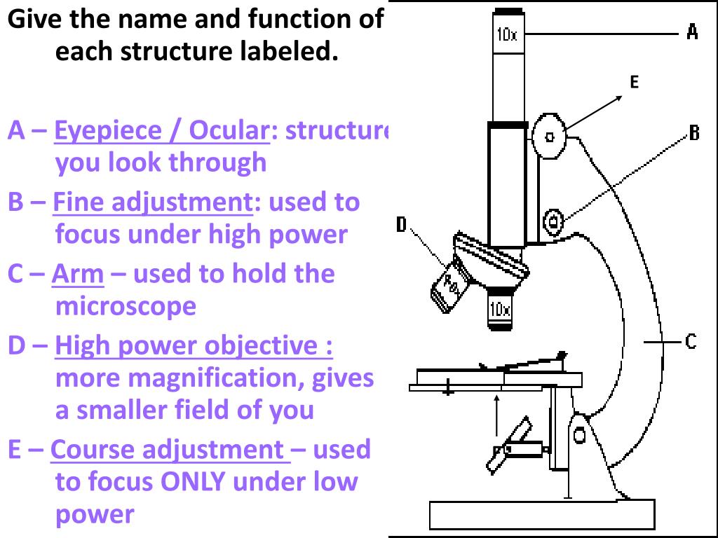
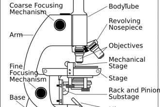
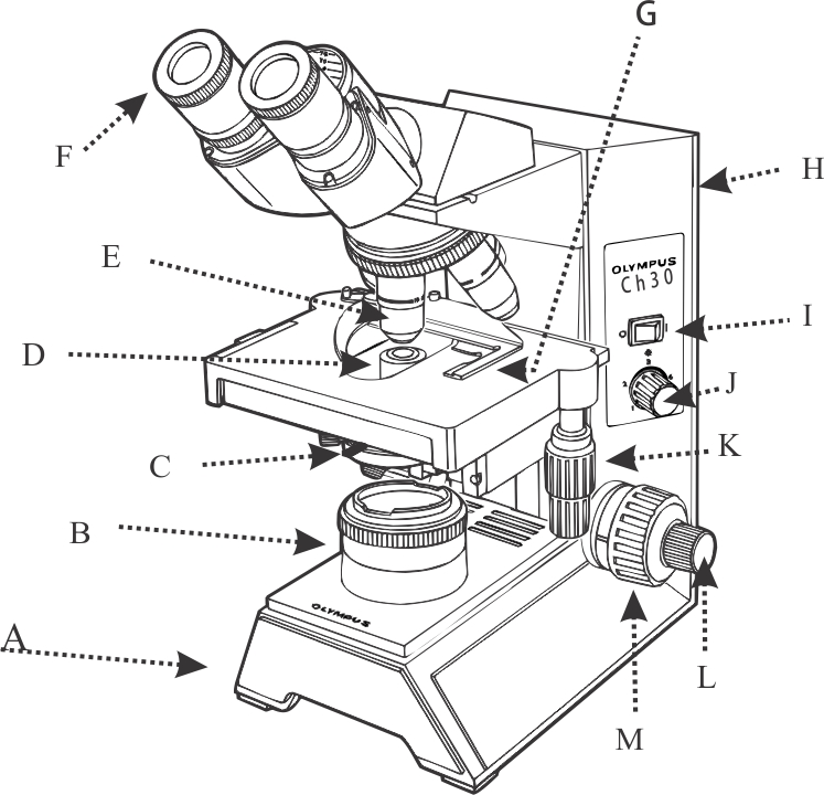
A physical wiring diagram for the human immune system | Nature 3.8.2022 · The human immune system is composed of a distributed network of cells circulating throughout the body, which must dynamically form physical associations and communicate using interactions between ... Compound Microscope Parts - Labeled Diagram and their Functions Labeled diagram of a compound microscope Major structural parts of a compound microscope There are three major structural parts of a compound microscope. The head includes the upper part of the microscope, which houses the most critical optical components, and the eyepiece tube of the microscope. Microscope Types (with labeled diagrams) and Functions Simple microscope labeled diagram Simple microscope functions It is used in industrial applications like: Watchmakers to assemble watches Cloth industry to count the number of threads or fibers in a cloth Jewelers to examine the finer parts of jewelry Miniature artists to examine and build their work Also used to inspect finer details on products Simple Microscope - Diagram (Parts labelled), Principle, Formula and Uses A simple microscope consists of Optical parts Mechanical parts Labeled Diagram of simple microscope parts Optical parts The optical parts of a simple microscope include Lens Mirror Eyepiece Lens A simple microscope uses biconvex lens to magnify the image of a specimen under focus.
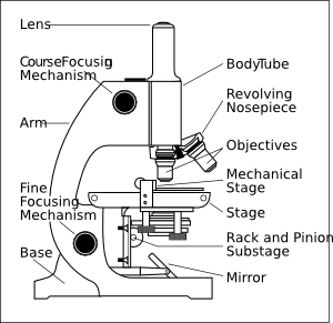
Autoclave - Wikipedia A medical autoclave is a device that uses steam to sterilize equipment and other objects. This means that all bacteria, viruses, fungi, and spores are inactivated. However, prions, such as those associated with Creutzfeldt–Jakob disease, and some toxins released by certain bacteria, such as Cereulide, may not be destroyed by autoclaving at the typical 134 °C for three minutes or 121 … Cells and Reproduction - BBC Bitesize The proper name for a living thing is a living organism. A living organism can be, amongst other things, a plant or an animal. Simple Microscope - Definition, Types, Working Principle & Formula A simple microscope consists of a convex lens of a short focal length. The below figure shows the ray diagram which subsequently forms the image of an object (or we can say a source of light). (Image will be Updated soon) F is the focal length of the lens. An object is placed between the focal length and the centre of the curvature. Parts of a microscope with functions and labeled diagram - Microbe Notes Figure: Diagram of parts of a microscope There are three structural parts of the microscope i.e. head, base, and arm. Head - This is also known as the body. It carries the optical parts in the upper part of the microscope. Base - It acts as microscopes support. It also carries microscopic illuminators.
Microscope Unlabelled Diagram - Micropedia
Labeling the Parts of the Microscope | Microscope World Resources Labeling the Parts of the Microscope This activity has been designed for use in homes and schools. Each microscope layout (both blank and the version with answers) are available as PDF downloads. You can view a more in-depth review of each part of the microscope here. Download the Label the Parts of the Microscope PDF printable version here.

Cell organelles | Cells: the basic units of life | Siyavula
Microscope Poster - Diagram with Labels | Teach Starter A poster containing a diagram with labels showing the key parts of a microscope. In Science it is important that students know how to use a variety of tools when conducting scientific experiments and inquiry. This poster focuses on the microscope and highlights its key parts. There are two print options available for this poster:

Simple Cuboidal
Lower Secondary Science LEARNER’S BOOK 8 - Issuu 22.2.2021 · Read Lower Secondary Science LEARNER’S BOOK 8 by Cambridge University Press Education on Issuu and browse thousands of other publications on our p...

11 Best Images of Cell Labeling Worksheet Answers - Cell Cycle and Mitosis Worksheet Answers ...
arXiv:2208.12069v1 [cond-mat.mes-hall] 25 Aug 2022 1 päivä sitten · eld microscope that can distinguish QD emission from back-re ected laser light by a cross-polarization scheme. Details on fabrication and measurement of the QDs can be found in Ref.6. An example of the observed spectra is shown in Fig. 1. Some general properties of the measured spectra can be described using the idealized diagrams shown in Fig. 2,

Pin on Diagrams/Charts/Maps
Microscope Poster - Diagram with Labels | Teach Starter A poster containing a diagram with labels showing the key parts of a microscope. Use this educational classroom poster in your science lessons to highlight the key parts of a microscope. A lot of equipment is used in science experiments and it is important to know the names of and understand each part of the equipment and how it works.

Labelled Diagram Of A Tick - Top Label Maker
Labelled Diagram of Compound Microscope The below mentioned article provides a labelled diagram of compound microscope. Part # 1. The Stand: The stand is made up of a heavy foot which carries a curved inclinable limb or arm bearing the body tube. The foot is generally horse shoe-shaped structure (Fig. 2) which rests on table top or any other surface on which the microscope in kept.

29 best Anatomy and Physiology images on Pinterest | Physiology, Anatomy and Anatomy reference
Simple Microscope Definition, Magnification, Parts And Uses - BYJUS Following are the parts of the simple microscope with their functions: Eyepiece: It is the lens that is used to study the samples and is placed at the top. It has a magnification of 10X to 15X. Base: This provides support to the microscope. Tube: This is used to connect the eyepiece to the objective lenses.
Microscope With Labels clip art (111146) Free SVG Download / 4 Vector
Microscope labeled diagram - slideshare.net Microscope labeled diagram 1. The Microscope Image courtesy of: Microscopehelp.com Basic rules to using the microscope 1. You should always carry a microscope with two hands, one on the arm and the other under the base. 2. You should always start on the lowest power objective lens and should always leave the microscope on the low power lens ...
Microscope labelling 11 - Teaching resources
GCSE Science: Required practical activities - AQA Using a light microscope to observe, draw and label cells in an onion skin . Materials . In addition to access to general laboratory equipment, each student needs: • a small piece of onion • a knife or scalpel • a white tile • forceps • a microscope slide • a coverslip • a microscope • iodine solution in a dropping bottle.
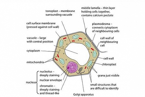
Plant cell Structure: Plant cell parts, Organelles and their functions and Diagram
Truth Giver of Humanity: What is the Human Race? - Blogger 6.5.2022 · Here is a diagram of melanin synthesis of black/brown and yellow pigment. The amino acid tyrosine is composed of the elements Carbon, Nitrogen, Oxygen, and Hydrogen as indicated by the diagram. However, the amino acid cysteine is composed of all these previous elements plus one more element called Sulfur as also indicated by the diagram.
1.1 Labelling Microscope - Labelled diagram
Microscope Labeling - The Biology Corner Students label the parts of the microscope in this photo of a basic laboratory light microscope. Can be used for practice or as a quiz. ... The type of microscope used in most science classes is the _____ microscope. 18. You should carry the microscope by the _____ and the _____. 19. The objectives are attached to what part of the microscope ...
Biology 521 Resources
16 Parts of a Compound Microscope: Diagrams and Video In compound microscopes with two eye pieces there are prisms contained in the body that will also split the beam of light to enable you to view the image through both eye pieces. 2. Arm The arm of the microscope is another structural piece. The arm connects the base of the microscope to the head/body of the microscope.
Foundations - Histology Epithelia and Skin - Embryology
Draw a neat labeled diagram for the formation of an image in a simple ... A simple microscope is a convex lens where in the image is kept between the focus of the lens so that the image formed is virtual and magnified. While drawing make sure the symmetry is taken into consideration. Complete step by step answer: Here is the labeled diagram outlining a simple microscope. To draw the ray diagram, place the object ...
Microscope Diagram With Labels And Functions - Micropedia
A Study of the Microscope and its Functions With a Labeled Diagram ... To better understand the structure and function of a microscope, we need to take a look at the labeled microscope diagrams of the compound and electron microscope. These diagrams clearly explain the functioning of the microscopes along with their respective parts. Man's curiosity has led to great inventions. The microscope is one of them.
Dissecting Microscope Labeled Diagram - Micropedia
Free Microscope Worksheets for Simple Science Fun for Your Students 1. Parts of a Microscope . The first worksheet labels the different parts of a microscope, including the base, slide holder, and condenser. If you have a microscope, compare and contrast this worksheet to it.Also, your kids can color this microscope diagram in and read the words to each part of the microscope.

Clipart Panda - Free Clipart Images
Microscope Parts and Functions Microscope Parts and Functions With Labeled Diagram and Functions How does a Compound Microscope Work?. Before exploring microscope parts and functions, you should probably understand that the compound light microscope is more complicated than just a microscope with more than one lens.. First, the purpose of a microscope is to magnify a small object or to magnify the fine details of a larger ...
Get Practice Labeling Parts Of A Microscope Gif - DirectScot
Microscope Labeling - The Biology Corner 1) Start with scanning (the shortest objective) and only use the COARSE knob . Once it is focused… 2) Switch to low power (medium) and only use the COARSE knob . You may need to recenter your slide. Once it is focused.. 3) Switch to high power (long objective).

The Microscope: Create a Labelled Diagram | Teaching Resources
Anatomy Chart - How to Make Medical Drawings and Illustrations Pathologic anatomy focuses on how diseases affect and change the human body. Histology studies microscopic anatomy such as tissues and cells visible only under a microscope. Anatomy charts serve two main purposes: education in the form of anatomy worksheets and presentation in the form of simple healthcare illustrations.
Post a Comment for "39 simple microscope diagram with labels"